Development of the lymphatic channels (Fig.11):
In the sixth week of pregnancy lymphatic vessels develop from lymph sacs which arise from the developing veins, which in turn are derived from mesoderm.
The first lymph sacs are the paired jugular lymph sacs at the junction of the internal jugular and subclavian veins. From the jugular lymph sacs, lymphatic capillary plexuses spread to the thorax, upper limbs, neck and head. Some of the lymphatic vessels are formed by enlargement of the plexuses in their respective regions. Each jugular lymph sac retains at least one connection with its jugular vein, the left one developing into the superior portion of the thoracic duct.
The next lymph sac is the unpaired retroperitoneal lymph sac, which is formed at the root of the mesentery of the intestine and develops from the primitive vena cava and mesonephric veins. Capillary plexuses and lymphatic vessels spread from the retroperitoneal lymph sac to the abdominal viscera and diaphragm. The sac establishes connections with the cisterna chyli but loses its connections with neighbouring veins.
The last of the lymph sacs are the paired posterior lymph sacs which develop from the iliac veins. The posterior lymph sacs produce capillary plexuses and lymphatic vessels of the abdominal wall, pelvic region and lower limbs. The posterior lymph sacs join the cisterna chyli and lose their connections with adjacent veins.
With the exception of the anterior part of the sac from which the cisterna chyli develops, all lymph sacs become invaded by mesenchymal cells and are converted into groups of lymph nodes.
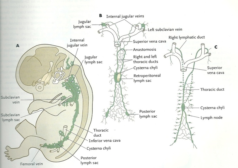
Figure.11: Development of lymphatic channels (Carlson, 2009).
Copyright © 2011 by Reem Bu Saeed.
Last updated: 30-07-2011.
Last updated: 30-07-2011.
