Fetal circulation (Fig.12):
Fetal circulation:
The placenta carries blood to the fetus through the umbilical vein. Half of the blood volume is delivered by the umbilical vein to the fetal ductus venosus, which carries the blood into the inferior vena cava. The other half of the blood volume is delivered by the umbilical vein to the liver through the inferior border of the liver in the right lobe by joining with the portal vein. Then the blood passes to the right atrium of the heart from the liver.
The foramen ovale, which is an opening between the right and left atria in the fetus, has an important role during fetal circulation. Most of the fetal blood flows through this foramen directly from the right atrium into the left atrium, consequently bypassing pulmonary circulation. After that, the blood flows into the left ventricle to pump the blood through the aorta to the entire body. From the aorta some blood moves through the internal iliac arteries to the umbilical arteries and re-enters the placenta.
There is another mechanism in fetal circulation. Some of the blood enters the right ventricle, which pumps the blood into the pulmonary artery. Because the fetus does not use its lungs yet for respiration, the ductus arteriosus connects the pulmonary artery and the aorta; this connection directs most of the fetal blood away from the lungs.
Fetal circulation changes at birth:
When the fetus is bornand with its first breath, the foramen ovale and ductus arteriosus close. The blood no longer bypasses the pulmonary circulation; as a result, the neonate’s blood becomes oxygenated and the systemic and pulmonary circulations start.
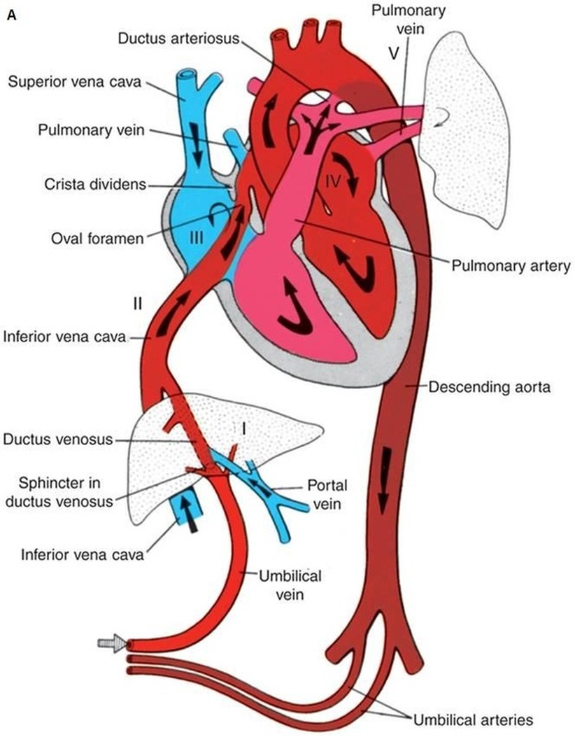
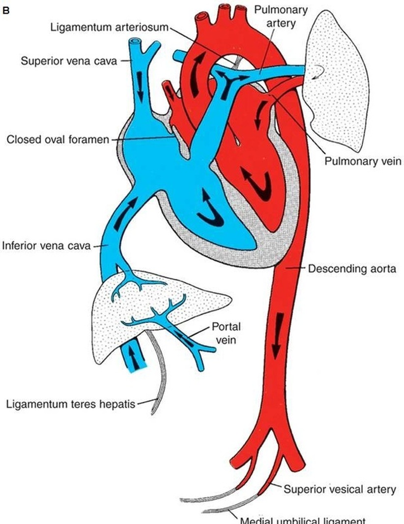
Figure.12: Fetal (A) and Neonatal (B) Circulation (Sadler, 2009).
<<< Back
Copyright © 2011 by Reem Bu Saeed.
Last updated: 30-07-2011.
Last updated: 30-07-2011.
