The aorta and its branches (Fig.11):
The ascending aorta arises from the aortic orifice at the base of the left ventricle in the middle mediastinum. The first branches from the aorta are the coronary arteries which arise from two sinuses associated with the aortic valve. The aorta then turns to the left to form the arch which gives off three branches that supply the upper limb, neck and head. The first and largest branch originates behind the manubrium of the sternum and is the brachiocephalic trunk. This trunk divides into two main arteries:
- Right common carotid artery which supplies the right side of the head and neck.
- Right subclavian artery which supplies the right upper limb.
The second branch is the left common carotid artery and is slightly posterior to the brachiocephalic artery. It ascends to the superior mediastinum along the left side of the trachea to supply the left side of the head and neck. The third branch is the left subclavian artery slightly posterior to the left common carotid artery and it also ascends up to the superior mediastinum along the left side of the trachea and supplies the left upper limb.
Other arteries arise from the underside of the aortic arch such as the bronchial and oesophageal arteries, which supply visceral structures within the thorax. The superior intercostal arteries arise from the brachiocephalic artery to supply the upper two intercostal spaces. The intercostal arteries supplying the nine lower intercostal spaces arise from the descending aorta.

Figure.11: The aorta and its branches.
The pulmonary trunk and its branches (Fig.12):
The pulmonary trunk arises at the junction of the confluence of the pulmonary arteries with the infundibulum of the right ventricle. The three sinuses of the pulmonary trunk also support the pulmonary valve and interdigitate with the fibrous extensions of the outflow tract in the semilunar attachments. As in the aorta, there is a marked sinutubular junction between the commencement of the ascending pulmonary trunk and the upper extent of each commissure of the valve leaflets. The trunk itself then divides into right and left pulmonary arteries, which extend to the hilum of their respective lung. In addition, there is an arterial ligament, a vestigial remnant of the ductus arteriosus, which conveyed deoxygenated blood to the aorta back to the placenta during foetal life. Finally, the pulmonary arteries themselves divide into several arteries to supply the bronchopulmonary segments of the right and left lung.
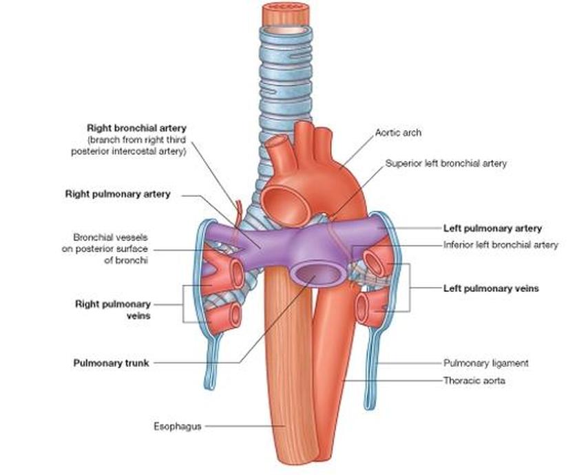
Figure.12: Pulmonary trunk and its branches (Drake et al, 2009).
Superior and inferior vena cava (Fig.13):
These two veins carry deoxygenated blood from the body to the right atrium in the heart. The superior vena cava arises at the junction of the right and left brachiocephalic veins at the lower edge of the right first costal cartilage. It terminates in the right atrium at the lower edge of the right third costal cartilage and carries deoxygenated blood from the upper half of the body. The inferior vena cava enters the right atrium at the lower right on the inferior aspect of the heart: it runs alongside the right side of the vertebral column. It carries deoxygenated blood from the lower half of the body.
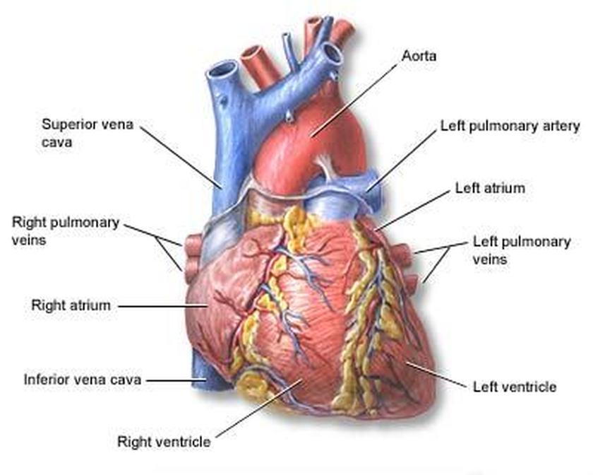
Figure.13: Superior and inferior vena cavae (Drake et al, 2009).
Copyright © 2011 by Reem Bu Saeed.
Last updated: 30-07-2011.
Last updated: 30-07-2011.
