Development of the arteries:
The aortic arches branch off from the aortic sac (Fig.8) . First, the blood exits from a common ventricle into a ventral aortic root from the pairs of aortic arches which distribute it through the branchial arches. The blood becomes reoxygenated through the gills when it passes from the aortic arch arteries into the capillary beds in gilled vertebrates. Then the blood passes from the aortic arches into paired dorsal aortae. Finally, the blood enters the regular systemic circulation.
During development, some arterial patterns become remodeled and some vessels regress. The first point is that there are six arches, numbered I,II,III,IV,V and VI with six pairs of arteries (in some references the fifth arch either never forms or forms incompletely and then regresses). Also, the aorticopulmonary septum divides the truncus arteriosus into ventral aorta and the pulmonary trunk. Then the right and left horns are formed by the aortic sac and lead to the brachiocephalic artery and proximal segment of the aortic arch.
On day 27, the first aortic arch disappears; however, there is a small portion that forms the maxillary artery. The second aortic arch also disappears, with the remaining portion forming hyoid and stapedial arteries. The third arch is large and the fourth and sixth are in the formation process. The primitive pulmonary artery is present.
On day 29, the first and second aortic arches disappear and the third, fourth and sixth arches are large. With further development, the third aortic arch forms the common carotid artery and first part of the internal carotid artery; the remainder of the latterartery is formed by the cranial portion of the dorsal aorta. The external carotid artery is considered to be a sprout of the third aortic arch. The fourth aortic arch forms part of the arch of the aorta, between the left common carotid and the subclavian arteries. In addition, it forms the most proximal part of the right subclavian artery. The fifth aortic arch never forms or forms incompletely and then regresses. The sixth aortic arch grows toward the developing lung bud.
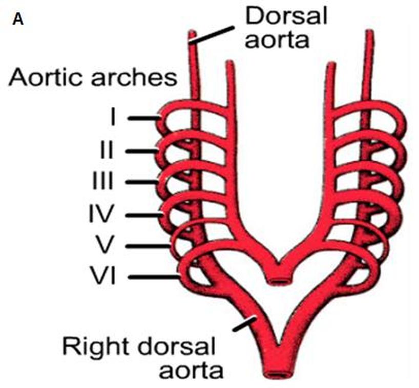
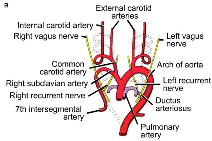
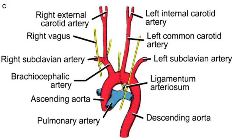
Figure.8: Formation of aortic arches (Sadler, 2009).
Major branches of the aorta:
The vitelline arteries (Fig.9): initially, the yolk sac is supplied by these arteries, then they fuse and form the arteries in the dorsal mesentery of the gut. In the adult, they are represented by the celiac, superior and inferior mesenteric arteries.
The umbilical arteries (Fig.9): initially, they form from the dorsal aorta as paired ventral branches. In the fourth week, each artery and its distal part obliterate and form the medial umbilical ligaments.
Coronary arteries: these grow out from the aorta, and then migrate and invade the aortic wall. They arise from the same cellular primordium as the future epicardium.
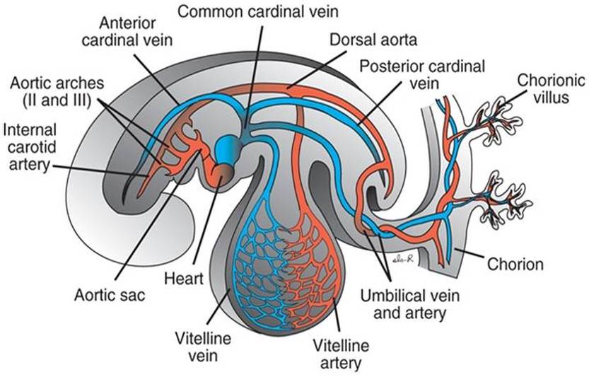
Figure.9: Formation of vitelline and umbilical arteries (Sadler, 2009).
Copyright © 2011 by Reem Bu Saeed.
Last updated: 30-07-2011.
Last updated: 30-07-2011.
